Blog
Cardiology AI in 2026: What's Actually Changed for Clinicians
🇬🇧 English edition - Auf Deutsch lesen (Link)
Where is AI in cardiology taking us? Here is where the field stands in mid-2026.

⏱️ Reading time: ~6 minutes
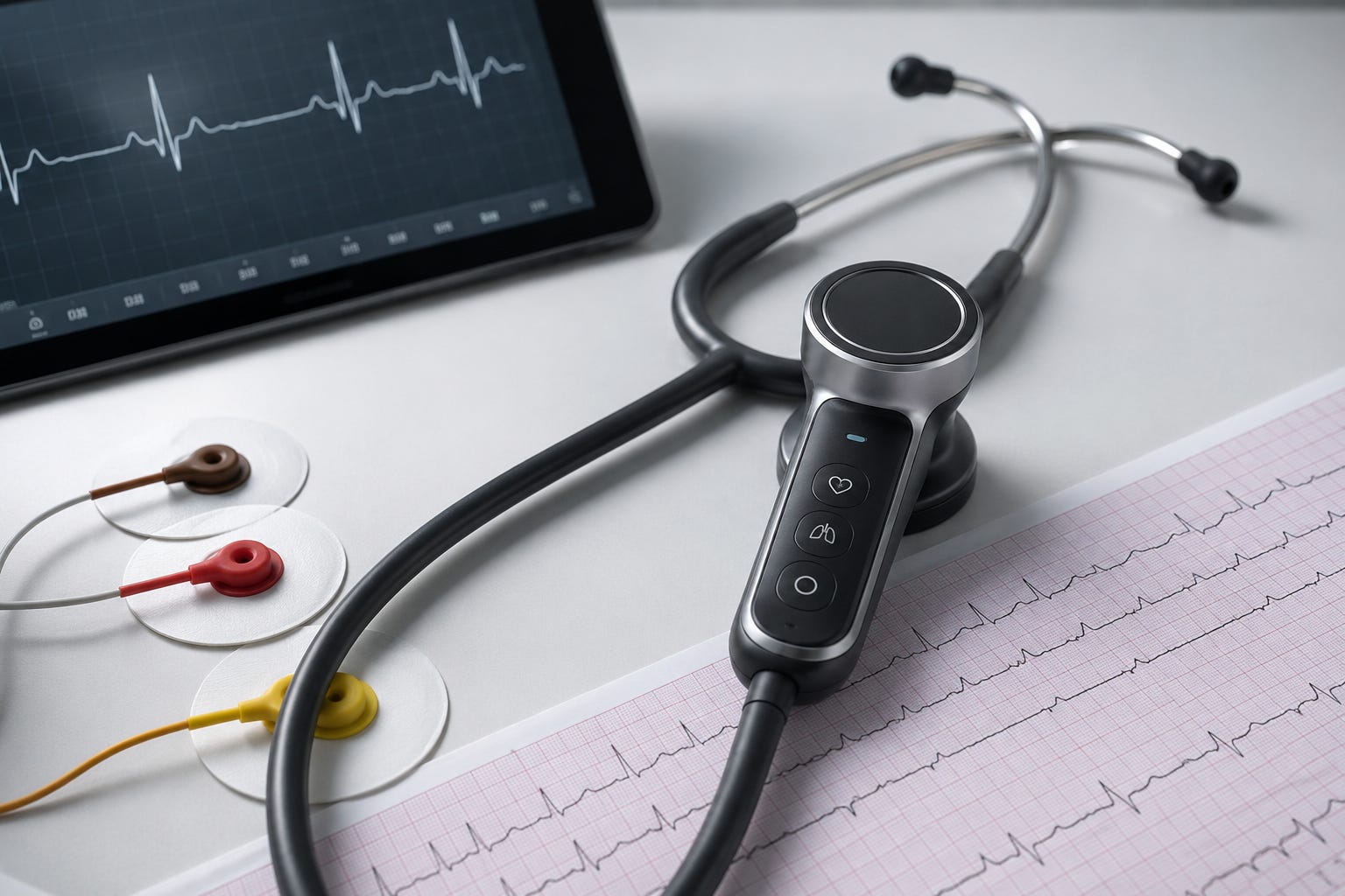
🧬 The ECG Is Learning to Detect Things It Never Could Before
Two AI algorithms from Anumana received FDA (US Food and Drug Administration) clearance in early 2026 for something a standard 12-lead ECG has never been validated to do before: detect cardiac amyloidosis and pulmonary hypertension (PH).[1]
Cardiac amyloidosis — in which amyloid protein deposits cause progressive myocardial stiffening and diastolic dysfunction — is chronically underdiagnosed; published registry data show a median diagnostic delay of over two years, with many patients waiting considerably longer.[15] PH carries a similar story: a median delay of two to three years from first symptoms to diagnosis.[16] Both algorithms flag patients who warrant further investigation and do not replace the imaging needed to confirm the diagnosis. Neither is yet available outside the US.
🚨 Chest Pain and the ECG: Catching What Standard Criteria Miss
A patient presents with chest pain. The ECG shows no ST elevation — by standard criteria, urgency appears lower. But the artery is occluded.
In an international study across European and US centres, an AI model identified occluded arteries from routine 12-lead ECGs with 80.6% sensitivity, compared to 32.5% using conventional STEMI (ST-elevation myocardial infarction) criteria.[2] Among patients labelled as NSTEMI (non-ST-elevation myocardial infarction), it flagged 61% of missed occlusions on the very first ECG.[2] Across the full study cohort, AI identified the diagnosis on average three hours earlier than standard criteria.[2]
A separate study of 1,490 patients with chest pain and no ST elevation found that AI achieved 99% specificity and a 98% negative predictive value (NPV) — meaning a positive result strongly confirms occlusion, and a negative result provides strong reassurance.[3]
PMcardio, the AI ECG tool used in several of these studies, is CE-marked as a Class IIb medical device under the EU Medical Device Regulation (MDR).[4] CE-marked tools can generally be used in Swiss hospitals once Swissmedic registration is completed.
⌚ Wearables Are Moving Beyond Rhythm Detection
Consumer wearables detecting atrial fibrillation (AFib) have been in clinical use for several years — that is no longer new. What is new is the scope of what these devices are now being validated to detect.
A study presented at the American Heart Association’s (AHA) 2025 Scientific Sessions trained an AI algorithm to identify structural heart disease — valvular disease, cardiomyopathy, and left ventricular hypertrophy (LVH) — from the single-lead ECG recorded by a smartwatch. The AI was developed using over 266,000 ECGs paired with echocardiograms and validated retrospectively in 44,591 adults across community hospitals and a population cohort. In a prospective study of 600 patients wearing a smartwatch on the same day as their echocardiogram, it correctly identified structural disease in 86% of cases and ruled it out with a 99% NPV.[6] These findings normally require echocardiography to detect. The smartwatch does not replace the echo — but it can identify which patients warrant one.
A related study demonstrated that a foundation model pre-trained on nearly 200,000 ECG-echocardiogram pairs could be fine-tuned for structural heart disease screening using as few as ~1,000 labelled ECGs — significantly lower than what conventional models require, and making deployment across different hospital settings more feasible.[7]
Kardia 12L (12-lead) is a portable five-electrode device that captures a standard 12-lead ECG outside of a hospital or clinic setting. Its AI received FDA clearance in 2026 for 39 cardiac determinations, including myocardial infarction (MI), axis deviations, and complex arrhythmias.[8] It is US-cleared only — no CE marking or Swissmedic registration yet.
The clinical implication is a shift in how to think about wearable data arriving from patients. The question is no longer only “is this AFib?” but increasingly “does this tracing suggest something structural that needs imaging?”
🫀 Digital Twins Guide Ablation for Ventricular Tachycardia
For patients with ventricular tachycardia (VT) that has not responded to medication, catheter ablation is the standard next step — but in post-infarction VT, recurrence is common, with conventional long-term success rates around 60%.[9]
The TWIN-VT trial, published in the New England Journal of Medicine in 2026, tested a different approach.[9] Before each procedure, a personalised digital twin of the patient’s heart was built from a contrast-enhanced cardiac MRI (magnetic resonance imaging) — a 3D computational model incorporating that patient’s specific scar tissue distribution and electrical architecture. Electrophysiologists could then simulate the arrhythmia circuit and identify the ablation target before the procedure began.
In ten patients with post-infarction VT, VT was non-inducible in all ten immediately after ablation. At 13 months, eight of ten remained free of recurrence without antiarrhythmic therapy.[9]
Ten patients is a small feasibility study, and the results are not yet practice-changing at scale. But the concept — planning a complex electrophysiology procedure on a patient-specific computational model before it begins — represents a meaningful shift in how AI is entering the procedural space.
🩻 AI in Cardiac Imaging: Echo and CT
Echocardiography
PanEcho is AI software that clinicians can use alongside a standard echocardiography workflow: the sonographer acquires the images as usual, and PanEcho simultaneously analyses the recordings across 18 diagnostic tasks — producing automated measurements and classifications without interrupting the acquisition process.
Trained on over one million echocardiographic videos and validated across 32,265 studies at four international cohorts — including point-of-care handheld devices — it detected moderate-to-severe left ventricular (LV) systolic dysfunction with an AUC (area under the receiver operating characteristic curve) of 0.98–0.99, and severe aortic stenosis with AUC up to 1.00.[10] Published in JAMA, this is one of the most comprehensively validated AI echocardiography systems to date. The point-of-care performance is particularly relevant for settings where expert echocardiographers are unavailable or overloaded.
Coronary CT
AI applied to coronary CT angiography (CCTA) is moving beyond simply measuring arterial stenosis. In a large international registry of over 3,500 patients, AI-based plaque characterisation — distinguishing stable calcified deposits from lipid-rich, high-risk plaques more prone to rupture — significantly improved prediction of major adverse cardiac events (MACE) beyond conventional risk scoring alone.[11] AI-assisted CCTA reports are increasingly including plaque vulnerability scores alongside stenosis measurements, and understanding what these mean is becoming a routine interpretive competency.
🩺 Not All That Glitters Is Gold
One of the largest real-world AI trials in clinical practice recently published its results. The TRICORDER trial ran across 205 NHS general practices in the UK, covering over 1.5 million registered patients.[5] It tested an AI-enabled stethoscope designed to improve detection of heart failure and arrhythmias.
Among patients actually examined with the device, heart failure detection nearly doubled and arrhythmia identification tripled compared with standard care.[5]
The finding that mattered more: adoption fell steadily throughout the trial. Clinicians reported the device added steps to already full appointments and did not connect to existing patient record systems.[5]
A tool that performs well in a controlled protocol but disrupts the real clinical encounter does not help real patients. Workflow integration is as important as diagnostic performance.
⚠️ What AI in Cardiology Still Cannot Prove
Most of what is described above has been validated on diagnostic accuracy — how well a tool detects a condition. Very few tools have yet shown that better detection translates into better patient outcomes: fewer myocardial infarctions (MI), lower mortality, improved quality of life.
The ESC (European Society of Cardiology) made this point explicitly in its 2025 roadmap on trustworthy AI, calling for outcomes evidence before these tools are embedded in clinical guidelines.[14] The AHA (American Heart Association) reaches the same conclusion: “few artificial intelligence tools have been shown to improve cardiovascular care sufficiently to be widely adopted.”[12]
🇨🇭 Where Switzerland Stands
Switzerland’s regulatory authority for medical devices is Swissmedic, which operates separately from the EU’s CE marking framework. A tool approved in the US or CE-marked in the EU requires its own Swissmedic registration before it can be used in Switzerland.
On reimbursement: as of mid-2026, no dedicated TARDOC position code for AI-enhanced cardiac diagnostics has been established.[17] AI interpretation is billed under the same codes as the underlying procedure — ECG, CT, or echocardiography. A dedicated reimbursement pathway is likely to follow, but does not yet exist. For which tools are actually in use at your institution, your cardiology department or clinical informatics team is a more reliable source than vendor websites.
📌 In Brief
ECG AI can now flag cardiac amyloidosis and pulmonary hypertension — both require imaging to confirm; neither algorithm is yet available outside the US
In chest pain without ST elevation, AI detects significantly more coronary occlusions than standard ECG criteria — CE-marked tools exist; check locally whether they are in use
Wearables are moving beyond AFib detection toward structural heart disease screening — validated at 86% sensitivity and 99% NPV in a prospective smartwatch study
PanEcho interprets 18 echocardiographic tasks simultaneously — validated at JAMA scale including on point-of-care devices, with direct implications for echocardiography lab workflow
Digital twin-guided VT ablation showed 100% immediate success in 10 patients — early feasibility data; a meaningful new direction for electrophysiology
Most cardiac AI has been validated on detection accuracy, not patient outcomes — the ESC and AHA have explicitly called for outcomes evidence before these tools enter clinical guidelines
📚 Sources
Anumana. FDA clearance for ECG-AI Pulmonary Hypertension Algorithm. BusinessWire, March 27, 2026. Link— FDA clearance for ECG-AI Cardiac Amyloidosis Algorithm. BusinessWire, April 7, 2026. Link
Herman R et al. International evaluation of an AI-powered ECG model detecting acute coronary occlusion MI. Eur Heart J Digit Health. 2024;5(2):123–133. Link
Nani F. AI-enabled ECG algorithm for detection of occlusive MI in suspected ACS without ST elevation. Presented at ESC Acute CardioVascular Care 2026. ESC Press Release
Powerful Medical. PMcardio CE marking under EU MDR. powerfulmedical.com
Kelshiker M et al. TRICORDER Trial. Imperial College London. 2026. Link
Aminorroaya A et al. AI tool detects structural heart disease using a smartwatch. AHA Scientific Sessions 2025. AHA Newsroom — Conference abstract; citation to be updated if published in a peer-reviewed journal
Knight E et al. Wearable-Echo-FM: an ECG echo foundation model for 1-lead electrocardiography. Eur Heart J Digit Health. 2026;7(4):ztag049. Link
AliveCor. FDA Clearance: Kardia 12L ECG System, 39 Cardiac Determinations. 2026. GlobeNewswire
Chrispin J et al. Digital Twin–Guided Ablation for Ventricular Tachycardia. N Engl J Med. 2026. Link
Holste G et al. Complete AI-Enabled Echocardiography Interpretation With Multitask Deep Learning. JAMA.2025;334(4):306–318. Link
van Rosendael A et al. AI-Driven Quantitative CCTA in Suspected Coronary Artery Disease: CONFIRM2 Registry.JACC Adv. 2026;5(3):102618. Link
Khera R et al. Use of Artificial Intelligence to Improve Outcomes in Heart Disease. Circulation. 2024. Link
Adedinsewo DA et al. AI-guided screening for cardiomyopathies in an obstetric population: a pragmatic RCT. Nat Med. 2024;30(10):2897–2906. Link
Asselbergs FW et al. Trustworthy AI in cardiovascular medicine: ESC roadmap. Eur Heart J. 2025;46(8):677. Link
Vogel N et al. Delays in diagnosis and treatment of ATTR cardiac amyloidosis: a real-world data analysis. ESC Heart Fail. 2025. Link
Didden AC et al. Time to diagnosis of pulmonary hypertension and diagnostic burden. Pulm Circ. 2023. Link
FMH. TARDOC Tarifbrowser — online tariff catalogue. tarifeambulant.fmh.ch
Liked this? Get new articles in your inbox.
Subscribe